Under the auspices of the respected Dean of the College of Dentistry, Prof. Dr. Raghad Abdul-Razzaq Muhammad Al-Hashimi
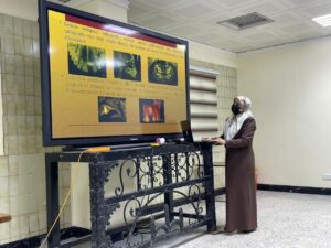
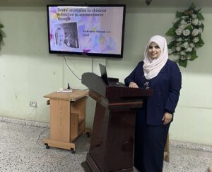
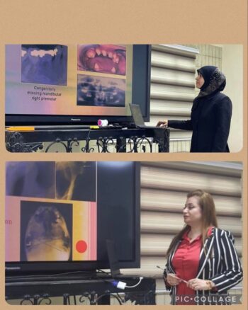
The College of Dentistry at the University of Baghdad organized a continuing education course entitled (Disorders of dental growth and its most important manifestations in radiographic imaging), in the presence of a number of faculty members in the college.
The course aimed to familiarize the participants with the definition of the most important types of dental growth disorders, their causes and effects, and the most important clinical changes that occurred and their manifestations in radiographic imaging.
The course included a full explanation of what growth disorders are, what are their most prominent symptoms, and how to diagnose them. Dental growth disorders may be due to abnormalities in the differentiation of the dental plate and tooth germs (abnormalities in number, size, and shape) or to abnormalities in the formation of hard dental tissues (abnormalities in structure). In some cases, both stages of differentiation are abnormal. Dental development disorders may be hereditary, acquired, or idiopathic. Not all dental developmental disorders are congenital.
The course included a number of topics:
The first axis: an introductory view of the tsunami variable virus
Discovered in early 2023, a new variant of Omicron called XBB.1.5, the most transmissible strain of the virus to date, is prevalent in the United States. Wear masks and take other mitigation measures. . They also watch over 300 Omicron offspring around the world.
The second axis: an explanation of the lack of tooth enamel formation, which is a rare disorder, and one of the biggest challenges that the doctor faces is complete rehabilitation with the formation of deficiency in tooth enamel. An early intervention plan and multidisciplinary treatment must be implemented for the indicated case while implementing careful evaluation, treatment planning, and careful surgical procedures In order to meet the aesthetic and functional requirements of the patient with modern technology.
The third axis: Describe the type of teeth, the number of teeth in deciduous and permanent teeth, and the appearance of teeth in both teeth on a large scale in previous research.
There are 20 teeth in the deciduous teeth and 32 in the permanent teeth. The teeth usually appear normal, although small differences may appear. Abnormal teeth can be detected through careful clinical or radiographic observation. Malformed teeth can have a negative impact on a person’s appearance as well as functions such as eating and can lead to psychological problems. They can be detected using various X-ray techniques, conventional and panoramic scans but cone beam computed tomography (CBCT) is the most accurate.
The fourth axis: describing the curvature of the teeth, where most scholars agree that there are two possible reasons for the curvature of the teeth: the first and most common cause is the occurrence of trauma on the corresponding deciduous tooth, which results in a distortion in the development of the permanent tooth. The bud is angular with the normal bud. The curvature of the teeth may manifest in many forms, including the absence of the affected tooth, the retention of deciduous teeth for long periods in the mouth, or the penetration of the apex from the oral cortical plate, or it may be asymptomatic…
The fifth axis: the types of changes that occur to the teeth of children with malignant tumors who are subjected to chemotherapy and radiotherapy in the stage of formation and growth of teeth, and I talked about the causes of these changes and their connection to the type of disease, the method and dose of treatment, and the age of the affected child, and dealt with the latest international studies published on this subject. Axis VI: The aim of the lecture is to provide an overview of the purposes of TMJ imaging modalities ranging from plain radiographs to more advanced ones that aim to assess the integrity and relationships of hard and soft tissues, confirm the extent or stage of progression of known disease, and evaluate the effects of treatment.
The course recommended that knowing the normal anatomy of the teeth is not only necessary in dentistry, but also in other specialties because various medical conditions can negatively affect the appearance, structure and position of the teeth. With a proper diagnosis, complications of abnormal teeth can be prevented.
Where complications can not only negatively affect the aesthetics of a smile, but it can also negatively affect speaking, biting, chewing, etc. In addition to reducing a person’s self-confidence, and in some cases leading to depression and social isolation.
The best options for the diagnostic imaging of abnormal teeth as well as the monitoring of treatment progress are OPG, CT scan and CBCT
